What I Treat › Cerebrovascular
Cerebrovascular
Moyamoya Disease
Moyamoya disease is a process where arteries in the brain narrow over time and eventually block off. The brain compensates by creating small blood vessels.
These new vessels are fragile and can rupture, causing bleeding in the brain. If they cannot supply enough blood, part of the brain can be damaged, which is a type of stroke.
Dr. Arias is Director of Neurovascular Surgery at HCA HealthONE Swedish
Symptoms
- Weakness or numbness on one side of the body
- Trouble speaking or understanding speech
- Mini-strokes (TIAs) that come and go
- In children, symptoms triggered by crying, exercise or heavy breathing
- Headaches
- Seizures
- Bleeding in the brain, more common in adults
How moyamoya disease is diagnosed
MRI and MR angiography
MRI shows any areas of past stroke, and MR angiography shows the narrowed arteries at the base of the brain.
Cerebral angiogram
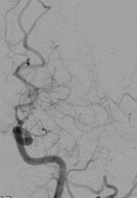
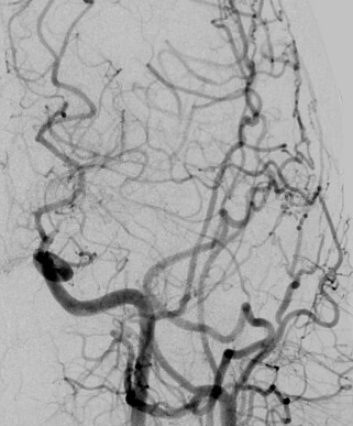
A catheter-based angiogram gives the clearest view of the narrowed arteries and the small replacement vessels, and is used to plan surgery.
Blood flow studies
Perfusion imaging measures how well blood is reaching each part of the brain, which helps decide whether and where bypass is needed.
Treatments for Moyamoya Disease in Denver & Fort Collins
Moyamoya tends to progress over time, so treatment focuses on restoring blood flow to the brain before a stroke happens.
Bypass Surgery
Surgery creates a new blood supply around the narrowed arteries, typically by redirecting an artery or muscle from the scalp to the brain. These are known as direct and indirect bypass techniques.
Medication
Medications such as aspirin are often used to lower the risk of stroke, alongside surgery or while monitoring.
Ongoing Monitoring
Regular imaging tracks blood flow to the brain over time, so changes can be caught and treated early.
Patient stories
26-year-old woman
26-year-old woman with left hemisphere Moyamoya disease, discovered after she developed right arm and leg weakness. She underwent a left indirect bypass and follow-up angiography performed 1 year after the procedure demonstrated marked increased vascularity in the left hemisphere.
More Cerebrovascular conditions
Request an Appointment with Dr. Arias
Dr. Eric J Arias, MD – Neurosurgeon & Neurointerventional Surgery