What I Treat › Cerebrovascular
Cerebrovascular
Arteriovenous Malformation (AVM)
An arteriovenous malformation (AVM) is a complex tangle of abnormal blood vessels in the brain that disrupts normal blood flow and oxygen circulation.
The vast majority of AVMs are present from birth but often go undetected until symptoms appear. Because they can cause bleeding, seizures and neurological damage, early diagnosis and treatment are essential.
Dr. Arias is Director of Neurovascular Surgery at HCA HealthONE Swedish
Symptoms
- Seizures
- Headaches
- Sudden severe headache, weakness or confusion from bleeding. Call 911
- Gradually worsening weakness or numbness
- Problems with speech, vision or balance
- No symptoms at all; some are found by chance
How an AVM is diagnosed
MRI and CT
These scans show the location and size of the AVM and any signs of past bleeding.
Cerebral angiogram
A catheter-based angiogram maps the arteries feeding the AVM and the veins draining it. This detailed picture is essential for choosing the safest treatment.
Treatments for Arteriovenous Malformation (AVM) in Denver & Fort Collins
If you or a loved one has been diagnosed with an AVM, a consultation can help you explore the options that fit you best. Early intervention is key to long-term outcomes.
Resection
Open surgery to remove the AVM, often recommended when it is accessible and has a high risk of bleeding. Recovery depends on its size and location, and many patients improve significantly.
Embolization
A minimally invasive catheter procedure that seals the AVM from within using a glue-like material. It can be used on its own or before surgery to lower the risk of bleeding.
Gamma Knife
Non-invasive, outpatient radiosurgery that targets the AVM without an incision. Best suited to small or deep AVMs, which gradually shrink and may disappear, with minimal effect on surrounding tissue.
Observation
Some AVMs that have never bled, especially in high-risk locations, may be monitored instead of treated, after a careful discussion of the risks of each approach.
Patient stories
31-year-old woman
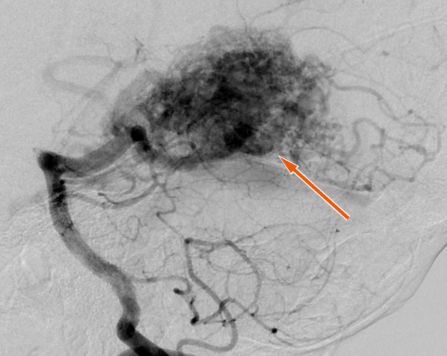
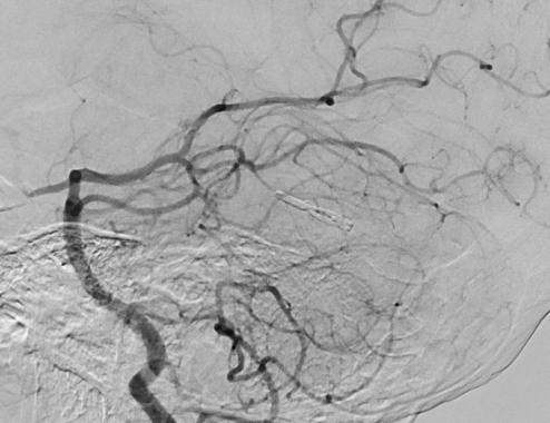
31-year-old woman with an arteriovenous malformation that was discovered after she had a seizure. She underwent gamma knife stereotactic radiosurgery and follow-up angiography performed 3 years after treatment demonstrated complete resolution of the malformation.
18-year-old man
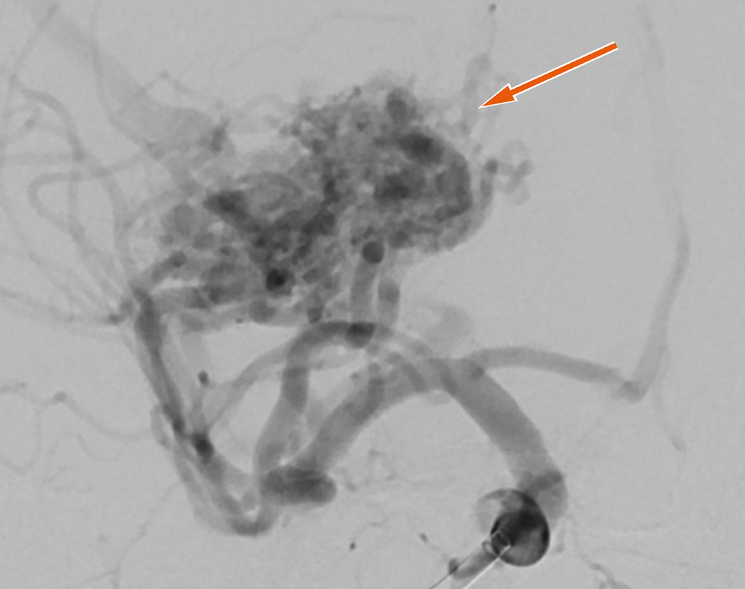
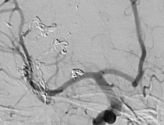
18-year-old man with a brain hemorrhage from a ruptured arteriovenous malformation. He underwent embolization and subsequent surgical resection of the lesion. We achieved removal of the entire lesion, and 6 months after his event, he is back in school.
More Cerebrovascular conditions
Request an Appointment with Dr. Arias
Dr. Eric J Arias, MD – Neurosurgeon & Neurointerventional Surgery